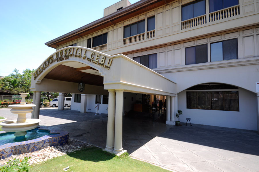
The Radiology Department of AMOSUP Seamen’s Hospital offers an array of diagnostic imaging procedures using modern facilities and skilled personnel that ensure reliable results for easy diagnosis of diseases while continually focussing on quality of patient care.
The radiologist consultants of the department are: Dr. Edwin Ferdinand C. Distor, Chairman; Dr Abigayl Lopez, CT-MRI Section Head, Dr Januario Paulito Tactacan, Interventional Radiologist; Dr Irene Bandong, Head-Breast Clinic, Dr Berlin Melissa Go, Dr Reginald Santos and Dr Marky Jod Pandes.
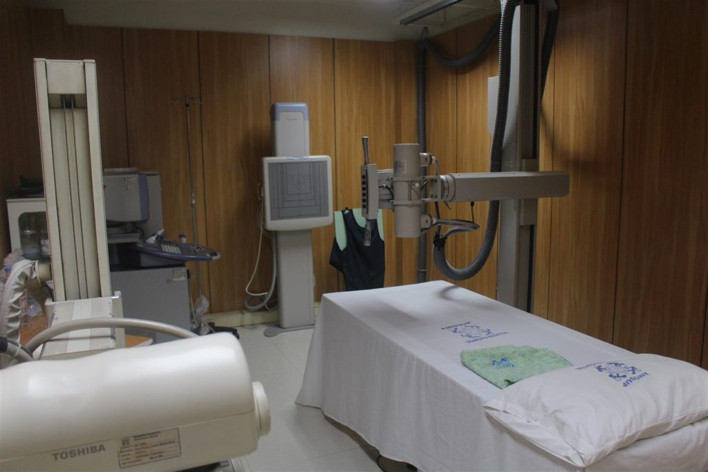
General Diagnostic X-ray
To keep pace with the advancing technology in diagnostic imaging, the hospital has shifted from screen film radiography (SFR) to digital radiography (DR) and acquired two digital x-ray imaging system and a computed radiography system for mobile radiography and mammography. The acquisition of these systems coupled with the PACS or picture archiving communication system has remarkably increased the quality and quantity of x-ray examinations done in the main section of the department.
In digital radiography the primary image recording is accomplished through the use of x-ray sensitive electronic detectors instead of phosphor screens or silver halide x-ray films. The image is displayed in digital format or printed on a film upon request. The quality of the image can be enhanced to improve the ability to detect diseases that will contribute to faster, easier diagnosis and greater treatment plan which means the patient will receive a better level of care. In digital radiography the x-ray images are instantly stored and ready to view in the computer which saves time resulting in greater productivity. The risk of losing important x-ray images is also eliminated because images are stored in the hard drive of the computer with a backup system.
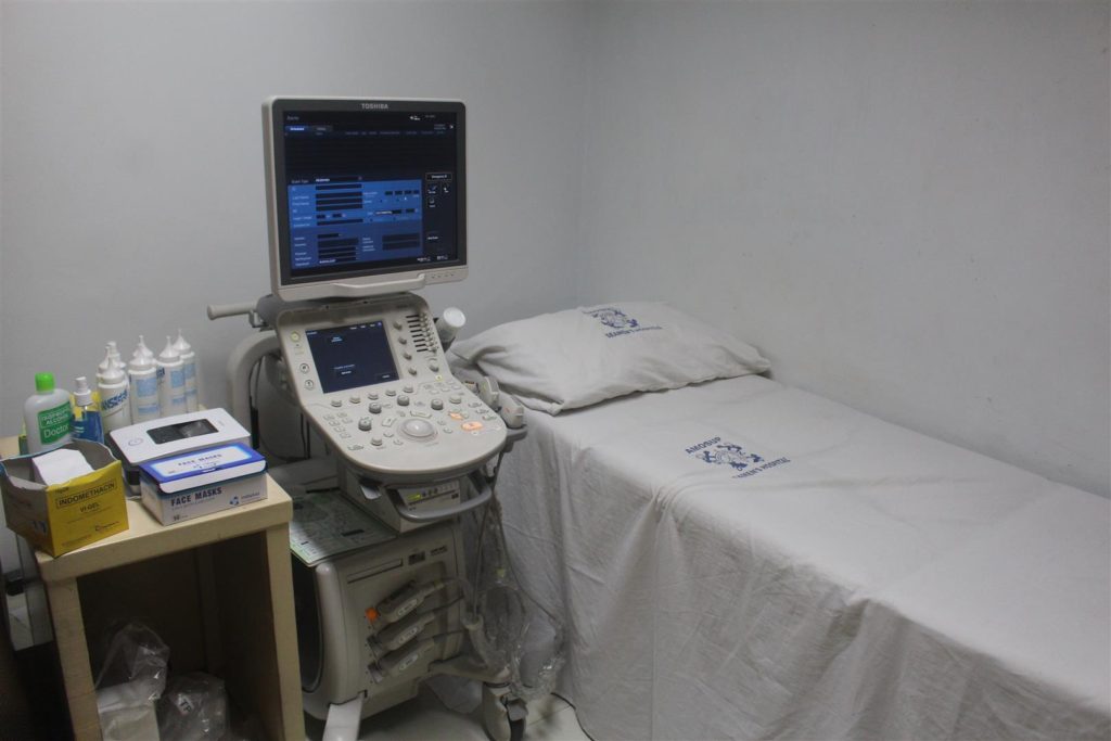
Ultrasound
Ultrasound, also called Sonography, is an imaging method using high frequency sound waves to produce images of structures within the body captured in real time. Unlike x-rays, there is no ionizing radiation associated with ultrasound imaging so it does not have the same risks as other imaging technique that uses ionizing radiation.
Abdominal ultrasound, which is done in the department, averages from 30-40 patients daily and the result released immediately after the examination.
Breast ultrasound is done to study breast tissues. It can help determine if an abnormality is solid which maybe a benign lump of tissue or a cancerous tumour; or fluid-filled such as a benign cyst; or both cystic and solid. Doppler ultrasound is done to assess blood supply in breasts lesions. The department has created a Breast Clinic section under Dr.Irene Bandong which is dedicated to the diagnostic study of the breast by means of a mammogram (x-ray) and breast ultrasound.
Vascular ultrasound also called a Duplex study is a non-invasive ultrasound method used to evaluate arteries and veins of the circulatory systems which includes the neck, abdomen, arms and legs to help identify blockages and blood clots. A Doppler ultrasound study is part of this technique to evaluate blood flow through a blood vessel. Dr.Marky Jod Pandes performs the procedure and makes the interpretation of the examination.
2D Echocardiography, commonly referred to as cardiac echo, is a sonogram of the heart using high-frequency sound waves and is routinely used in diagnosis, management of patients with any suspected or known heart diseases. A certified trained 2D Echo technician of the department, performs the procedure while a Cardiologist makes the interpretation.
Interventional Radiology is a subspecialty of radiology encompassing procedures performed using imaging guidance such as fluoroscopy, CT scan and ultrasound to diagnose and treat a wide variety of medical conditions. The most common procedures done at the department are CT and Ultrasound guided biopsies and ultrasound guided needle placement.
Extracorporeal shock wave lithotripsy (ESWL) is a technique for treating kidney stones and ureteral stones that does not require surgery. High energy shock waves are passed through the body to pulverise the stone so that they can pass from the body along with the urine. It takes 30 mins to 45 mins to complete the treatment. After treatment, some patients may still have stone fragments that are too large to be passed as revealed in the ultrasound exam. Treatment may be repeated if symptoms persists. Although this procedure is under the surgery department, a trained radiologic technologist performs the procedure under the supervision of the attending physician.
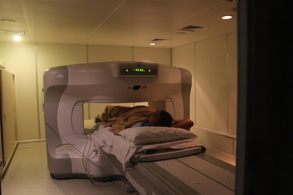
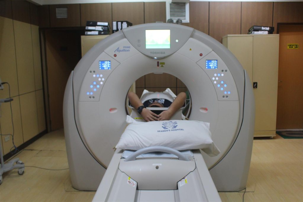
CT Scan and MRI Section. The CT scan section of the department has continued to keep pace with advancing technology with the acquisition of the 160 slice Toshiba CT Aquilon Prime, speeding up workflow and reducing exposure dose to the patient. Equipped with cutting edge technology for dose reduction and gantry bore of 78cm, the innovative CT Aquilon prime defines the next generation of CT Scanners. The average number of procedures done was increased and the results released within 24-48 hours after the completion of the examination. MRI examination of the head, neck, spine, shoulder and knees with and without contrast media are also performed.
In 2016, the Radiology Department served a total of 25,550 in all its sections, lower than the 28,247 recorded in 2015.The department under the dynamic leadership of its chairman Dr Edwin Ferdinand C. Distor will continue to keep the mission of the department “to provide and sustain quality patient care in the medical imaging field” and support AMOSUP Seamen’s Hospital’s vision of becoming a leading recognised maritime medical cenrtre. SF